YOUR DOG'S HEALTH


PennHIP is a not-for-profit veterinary health service at the University of Pennsylvania.
PennHIP is a multifaceted radiographic
screening method for hip evaluation. The technique assesses the
quality of the canine hip and quantitatively measures canine hip
joint laxity. The PennHIP method of evaluation is more accurate
than the current standard in its ability to predict the onset of
osteoarthritis (OA). Osteoarthritis, also known as degenerative
joint disease (DJD), is the hallmark of hip dysplasia (HD).
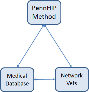
PennHIP is more than just a radiographic technique. It is also a
network of veterinarians trained to perform the PennHIP
methodology properly and, perhaps most importantly, it is a
large scientific database that houses the PennHIP data.
Radiographs are made by certified PennHIP members worldwide and
are sent to the PennHIP Analysis Center for evaluation. The
resulting data is stored in the database, which is continually
monitored as it expands. As more information becomes available,
the PennHIP laboratory is able to obtain more precise answers to
questions about the etiology, prediction and genetic basis of
hip dysplasia.
PennHIP publishes its findings in scientific journals. Published
information is disseminated to all PennHIP members; it is also
shared with interested breed clubs and routinely appears in
publications within the dog fancy.
PennHIP is composed of three major components:
The
PennHIP method is a novel way to assess, measure and interpret
hip joint laxity. It
consists of three separate radiographs: the distraction
view, the compression
view and the hip-extended
view. The distraction view and compression view are
used to obtain accurate and precise measurements of joint laxity
and congruity. The hip-extended view is used to obtain
supplementary information regarding the existence of
osteoarthritis (OA) of the hip joint. (The hip-extended
view is the conventional radiographic view used to evaluate the
integrity of the canine hip joint.) The PennHIP technique is
more accurate than the current standard, and it has been shown
to be a better predictor for the onset of OA.
The radiographs pictured here are of the same
dog, yet the hip joint laxties in each view look
very different. Notice that the hips in the distraction view
appear to be much looser than they do in the hip-extended view.
| Distraction View | Compression View | Hip-Extended View |
|
|
 |
 |
|
The obvious contrast in joint laxity
between the distraction and hip-extended views
demonstrates the fundamental difference between the two
radiographs. The
looser the joint on the distraction view, the greater is
the chance that the hip will develop OA.
The hip-extended view tends to mask true hip joint
laxity because the joint capsule iswound up into
a tightened orientation when the hips are extended. This
explains why measurable joint laxity on the distraction
view is always greater than the measurable laxity from
the hip-extended view. In fact, distraction laxity is up
to 11 times greater depending on the breed of dog under
study. The compression view is used to determine the "goodness of fit" of the femoral heads into the acetabula. In a hip with OA, the remodeling that occurs in the acetabulum and/or the femoral head, will often result in an ill-fitting "ball" and "socket". |
||
In 1983, Dr. Gail Smith from the University of Pennsylvania School of Veterinary Medicine conceived and developed a new scientific method for the early diagnosis of CHD. Research conducted in his laboratory proved the diagnostic method to be capable of estimating the susceptibility for CHD in dogs as young as sixteen weeks of age. In 1993, Dr. Smith established PennHIP, a cooperative scientific initiative, to serve as a multi-center clinical trial of the new hip dysplasia diagnostic technology. The program was successful and quickly grew beyond the capacity and purpose of a university research laboratory. Initially, the University of Pennsylvania licensed PennHIP to outside biotech companies in order to make the technology available for widespread public use and to allow Dr. Smith and his colleagues to continue their research at the School of Veterinary Medicine. PennHIP has recently been reacquired by the University of Pennsylvania and is now a not-for-profit organization.